Powered By


 Continue with Facebook
Continue with Email
Continue with Facebook
Continue with Email
Doctors frequently use X-rays during the diagnosis of lung cancer. These imaging tests help your cancer care team see within the lungs and detect various problems, such as unusual structures, tissue changes, or other signs of disease.
X-rays use radiation to visualize the tissues in your body. Lung tumors and other masses can often be seen on X-ray. Some other imaging tests, such as computed tomography (CT) scans and positron emission tomography (PET) scans, yield clearer pictures but are more expensive and time-consuming.
Taking chest X-rays often involves getting multiple images from two different angles. You may need an X-ray from the front or back, and one from the side. During the process, an X-ray technologist will use shields to cover any parts of your body that aren’t being X-rayed, help you move into the correct position, and take the images. Later, the images will be reviewed by a radiologist, a doctor who specializes in imaging tests.
Doctors typically use X-rays to help diagnose lung cancer. In some cases, they’ll also use X-rays to monitor whether treatments are working or to screen for lung cancer in people who are at risk of developing this condition. However, other imaging tests are more often used in these situations.
If your doctor thinks there’s a chance that you have lung cancer or another lung problem, they will usually recommend a chest X-ray as the first test. If the X-ray indicates that there’s a mass, lesion, or abnormal tissue in the lung, your doctor may have you undergo additional imaging tests, such as a CT, MRI, or PET scan. These tests can provide further information about abnormalities in the lung.
In order to make an official lung cancer diagnosis, doctors need to look at lung tissue samples to see whether there are cancerous cells. To do this, they may:
Lung cancer treatments may successfully shrink or eliminate lung tumors. However, there is a chance that the cancer can relapse (come back). Because of this, people with lung cancer need follow-up care to look for signs that the cancer has returned.
Some past research has found that once a person completes their treatment, getting regular chest X-rays can help detect relapsed tumors. However, other research has found that CT scans are more effective than X-rays at catching relapsed lung cancer in its early stages. The American Society of Clinical Oncology recommends that people with stage 1, 2, or 3 non-small cell lung cancer (NSCLC) get a chest CT scan every six months after completing treatment. These scans should continue for two years.
Lung cancer screening tests look for signs of lung cancer in people who are at risk for developing this condition. Experts recommend yearly lung cancer screening for current or former smokers between the ages of 50 and 80.
Past research has analyzed whether chest X-rays can be used as a lung cancer screening tool. However, CT scans are better at detecting tumors in the lungs. When they are used for screenings, they can more effectively reduce the chances that a person will die from lung cancer. Health professionals generally recommend low-dose CT scans for lung cancer screening.
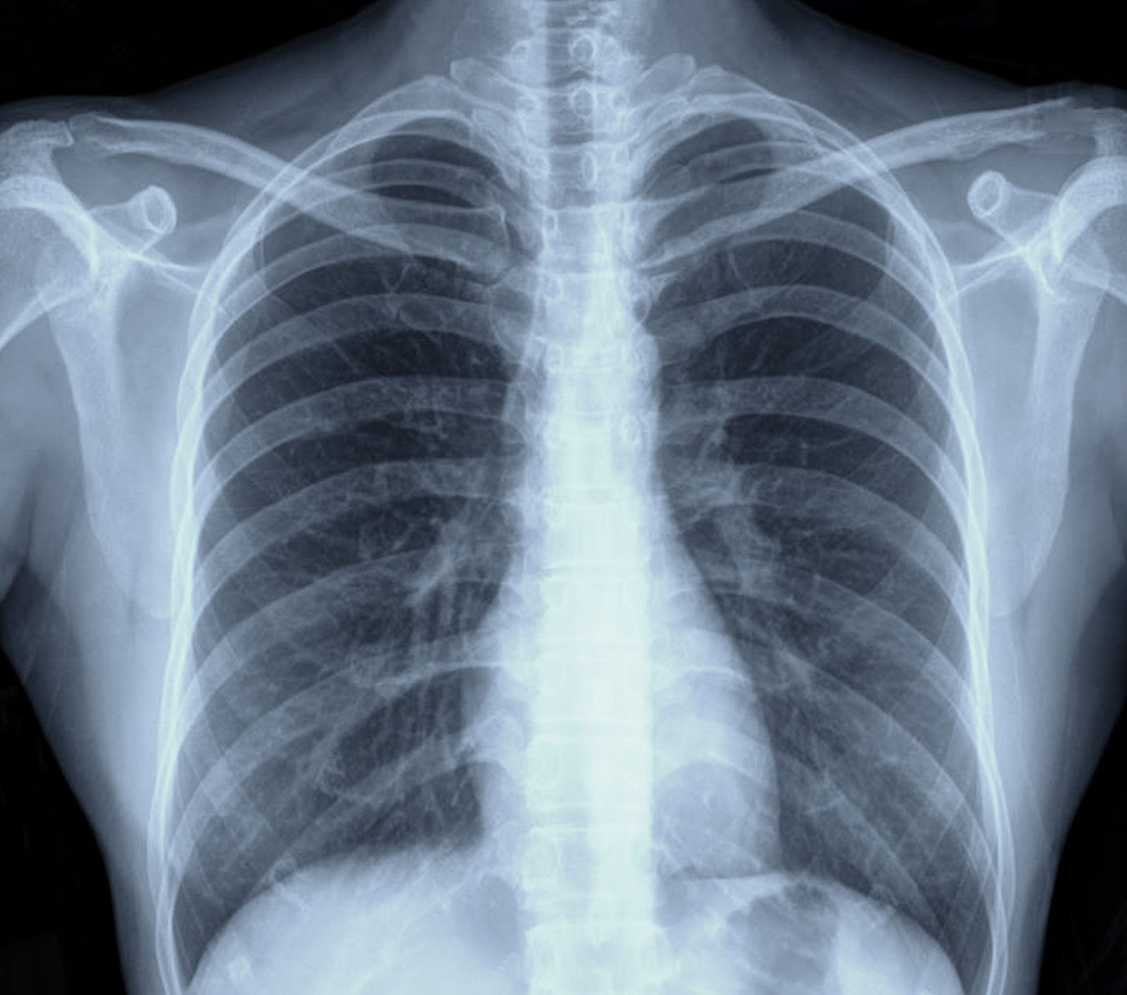
X-rays produce black-and-white images called radiographs. When you have an X-ray taken, a machine is placed on one side of your body. It emits a beam of radiation that travels through the tissues of your body and hits a detector or film positioned on the opposite side of your body. Softer tissues don’t interfere very much with the beam of radiation and allow most of it to travel through your body, leading to darker gray areas on the final X-ray image. On the other hand, denser tissues block more of the radiation from passing through, leading to white or light gray areas.
In a chest X-ray, certain tissues will appear in characteristic ways:
An X-ray can display the general size and location of a lung tumor. It may provide a clue as to whether the tumor has begun to grow into surrounding tissues. However, more detailed imaging tests, such as CT scans, are needed to get an accurate view of the tumor’s size and shape as well as the cancer stage (how far within the body the cancer has spread).
The way that a tumor appears on an X-ray may vary slightly based on the lung cancer type. Although chest X-rays can provide small clues as to which type of lung cancer a person may have, doctors won’t know for sure until other diagnostic tests are performed.
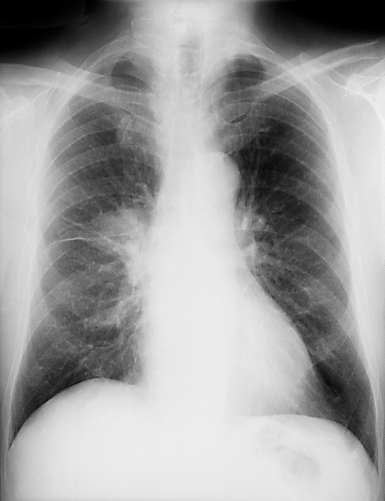
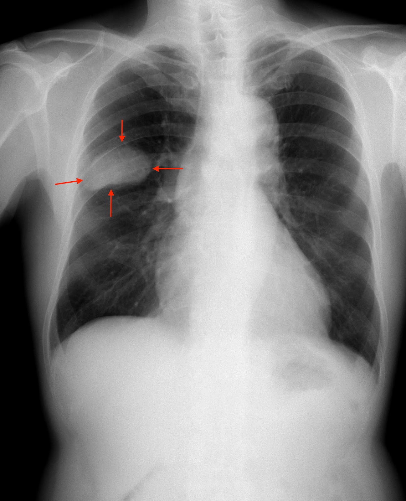
The following images represent a small sample of ways cancer can present on a chest X-ray. They are meant to give you a sense of what cancer might look like so you have a general understanding before your physician explains the specifics of your own images.
There are several types of NSCLC, which can vary in size and appear in different places within the lungs.
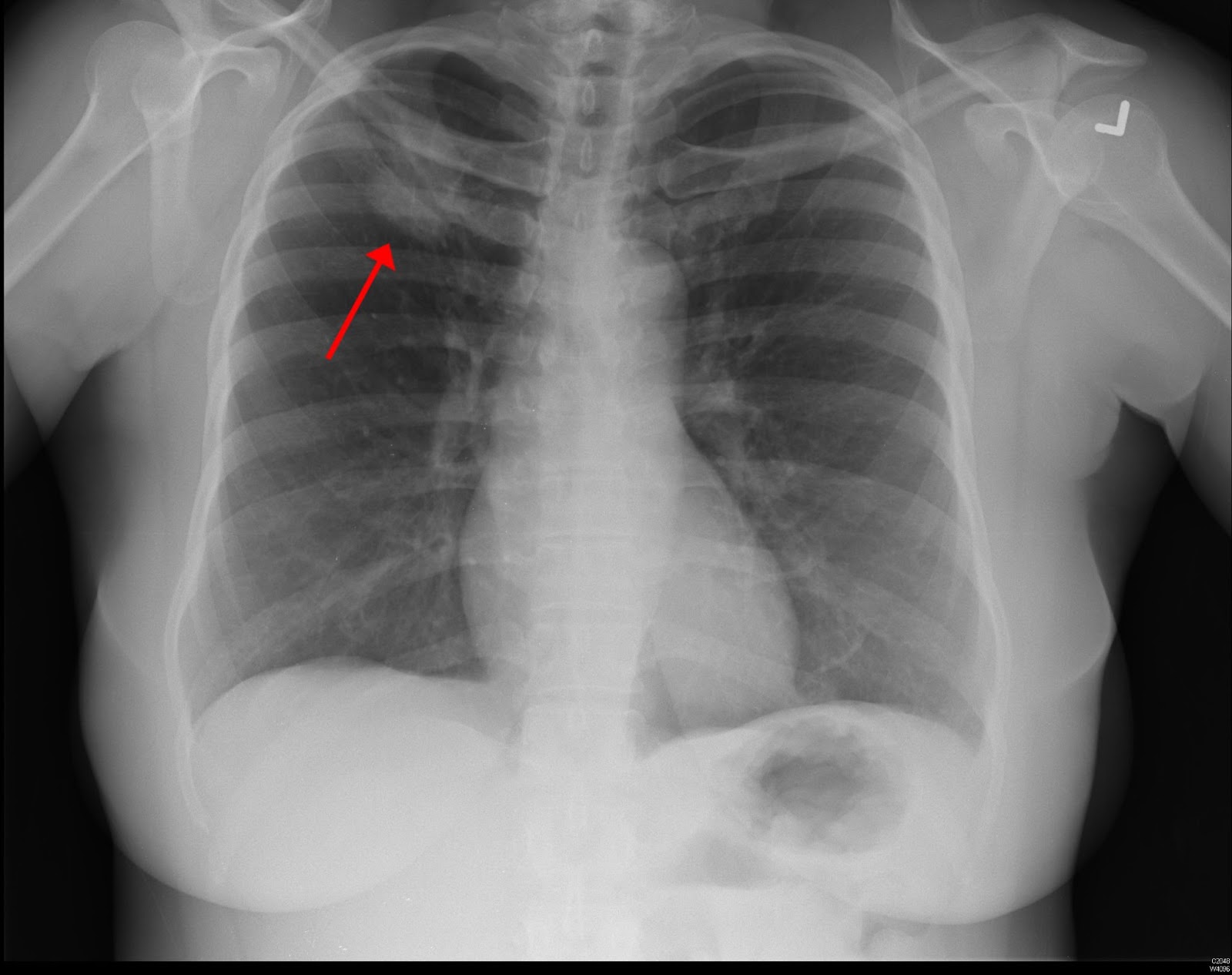
This type of NSCLC is often located on the outer surface of the lungs. For about half of people with adenocarcinoma, the chest X-ray will show that the tumor is starting to grow into lung tissues or other tissues located toward the middle of the chest.
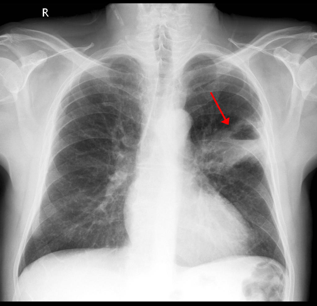
Squamous cell carcinoma tumors are often found toward the middle of the lung. Many squamous cell carcinomas have cavitation — they contain one or more air pockets that may show up on an X-ray as a dark spot surrounded by lighter-colored tissue.
A lung segment or lobe (a small part of the lung) collapses in many people with squamous cell carcinomas. This means that air leaks out of the lung into the surrounding space in the chest. Because there’s less air, the collapsed segment or lobe may show up as lighter gray on an X-ray.
These rare tumors are often located in the middle of the lung. Up to one-third of carcinoid tumors contain calcification (small areas of calcium), which can show up on an X-ray as small white dots.
SCLC tumors often appear toward the middle of the chest.
It’s important to know, these images are just some of many ways lung cancer may appear on a chest X-ray. A chest X-ray alone is likely not enough to rule out lung cancer, especially if you are experiencing symptoms. You can ask your health care team to help you understand your radiology images. The images shown in this article give you a general idea of what’s abnormal, but only a trained medical professional can tell you for sure what’s normal and abnormal.
MyLungCancerTeam is the social network for people with lung cancer and their loved ones. On MyLungCancerTeam, more than 12,000 members come together to ask questions, give advice, and share their stories with others who understand life with lung cancer.
Have you received chest X-ray as part of screening, diagnosis, or ongoing monitoring of lung cancer? Share your experience in the comments below, or start a conversation by posting on your Activities page.
Get updates directly to your inbox.


 Continue with Facebook
Continue with Email
Continue with Facebook
Continue with Email
Continue with Facebook
Continue with Email
Continue with Facebook
Continue with Email
Become a member to get even more


 Join
Join


 Your Privacy Choices

This is a member-feature!
Sign up for free to view article comments.
Does this look abnormal? I have other photos as well, if had double pneumonia, asma, smoker 20 yrs
Pbtype 1-2 tachardia 120 resting morning before anything
Currently CNA experienced chest and rib… read more
We'd love to hear from you! Please share your name and email to post and read comments.
You'll also get the latest articles directly to your inbox.